Health and MedicineDiagnostic Equipment, Tests, and Techniques |
What is nuclear magnetic resonance imaging? |
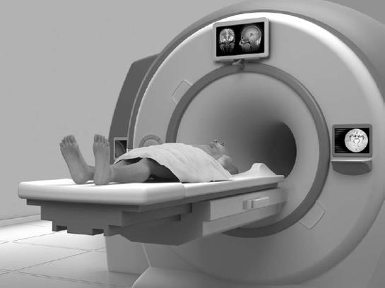
Magnetic resonance imaging (MRI), sometimes called nuclear magnetic resonance imaging (NMR), is a non-invasive, non-ionizing diagnostic technique. It is useful in detecting small tumors, blocked blood vessels, or damaged vertebral disks. Because it does not involve the use of radiation, it can often be used where X rays are dangerous. Large magnets beam energy through the body causing hydrogen atoms in the body to resonate. This produces energy in the form of tiny electrical signals. A computer detects these signals, which vary in different parts of the body and according to whether an organ is healthy or not. The variation enables a picture to be produced on a screen and interpreted by a medical specialist.
The concept of using MRI to detect tumors in patients was proposed by Raymond Damadian (1936–) in a 1972 patent application. The fundamental MRI imaging concept used in all present-day MRI instruments was proposed by Paul Lauterbar (1929–2007) in an article published in Nature in 1973. Lauterbur and Peter Mansfield (1933–) were awarded the Nobel Prize in Physiology or Medicine in 2003 for their discoveries concerning magnetic resonance imaging. The main advantages of MRI are that it not only gives superior images of soft tissues (like organs), but can also measure dynamic physiological changes in a non-invasive manner (without penetrating the body in any way). A disadvantage of MRI is that it cannot be used for every patient. For example, patients with implants, pacemakers, or cerebral aneurysm clips made of metal cannot be examined using MRI because the machine’s magnet could potentially move these objects within the body, causing damage.