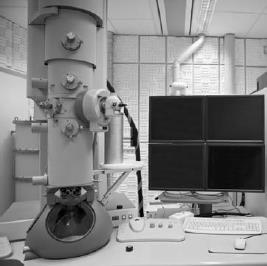
The theoretical and practical limits to the use of the optical microscope were set by the wavelength of light. When the oscilloscope was developed, it was realized that cathode-ray beams could be used to resolve much finer detail because their wavelength was so much shorter than that of light. In 1928 Ernst Ruska (1906–1988) and Max Knoll (1897–1969), using magnetic fields to “focus” electrons in a cathode-ray beam, produced a crude instrument that gave a magnification of 17, and by 1932 they had developed an electron microscope having a magnification of 400. By 1937 James Hillier (1915–2007) had advanced this magnification to 7,000. The 1939 instrument Vladimir Zworykin (1889–1982) developed gave 50 times more detail than any optical microscope ever could, with a magnification up to two million. The electron microscope revolutionized biological research: for the first time scientists could see the molecules of cell structures, proteins, and viruses.