Muscular SystemMuscle Structure |
What causes muscular dystrophy? |
Often thought of as a single disorder, muscular dystrophy is a group of genetic diseases characterized by progressive weakness and degeneration of the skeletal muscles that control movement. Missing or abnormal dystrophin causes muscular dystrophies. There are 30 types of muscular dystrophies and they are subdivided by mode of inheritance, age of onset, and clinical features. Discovery of the gene that causes the most common forms of muscular dystrophy took many years because dystrophin compromises only 0.002 percent of the protein in skeletal muscle. There is no specific treatment for muscular dystrophies. Ultimately, fat and connective tissue replace muscle.
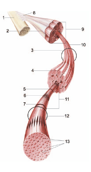
Muscle fiber. 1 = Periosteum: A tough, fibrous connective tissue that covers the surface of bones, rich in sensory nerves, responsible for healing fractures; 2 = Epimysium: Fibrous tissue enveloping the entire muscle and continuous with the tendon; 3 = Fascicle: a group of fibers that have been bound by perimysium; 4 = Endomysium: a delicate connective tissue that surrounds each muscle fiber; 5 = Z-line: boundary of two sarcomeres; 6 = H-zone: consists of stacks of myosin filaments; 7 = Z-line; 8 = Tendon: a dense, fibrous connective tissue that is continuous with the periosteum and attaches muscle to the bone; 9 = Belly: thick contractile portion (or body) of the muscle; 10 = Perimysium: fibrous tissue that extends inward from epimysium, surrounding bundles of muscle. Each bundle bound by perimysium is called a fascicle; 11 = Sarcomere: portion of muscle fibers found between two Z-lines; 12 = Muscle fibers: long, cylindrical, multinucleated cells with striations; 13 = Myosin actin: thick and thin filaments active during muscle contraction. Anatomical Chart Co.